Pelvic Floor Mri Protocol
Https Www Abdominalradiology Org Resource Resmgr Education Dfp Pelvicfloor Mri Imaging Mri Of Pelvic Floor Dysfunct Pdf

Position Liver Mri Liver Pancreas


Gynaecologic Mri Pelvis Uterus Cervix And Adnexal Protocols And Planning Indications For Mri Female Pelvis
Mr Female Pelvis Soft Tissue Pelvis W Wo Body Protocol Ohsu
Getting Ready For An Mri Of Your Pelvis Sansum Clinic

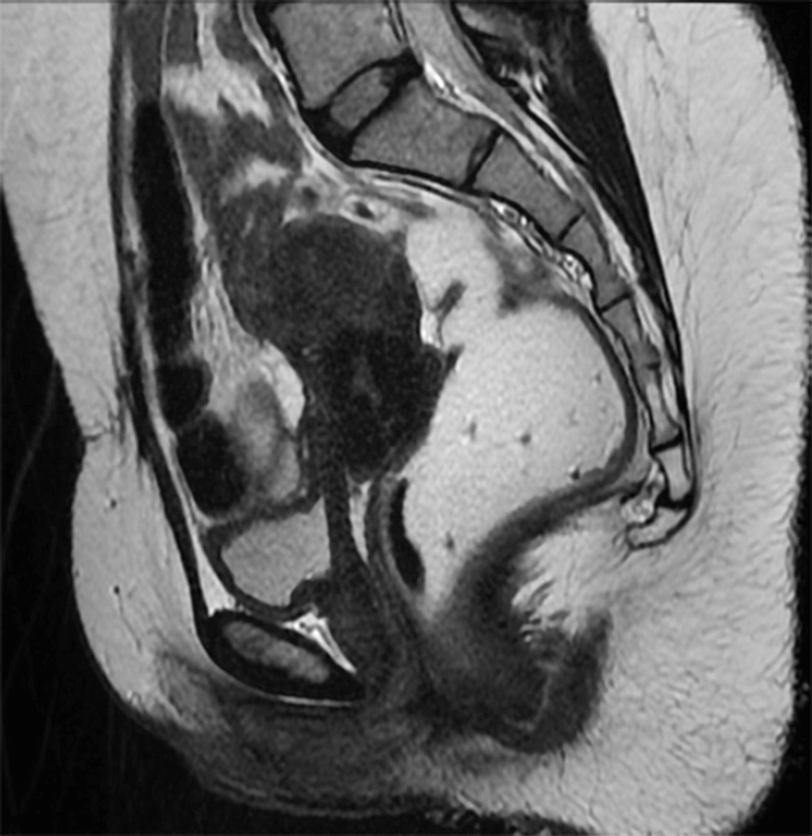
Dynamic pelvic floor magnetic resonance imaging mri is a noninvasive test that uses a powerful magnetic field radio waves and a computer to produce detailed pictures of the pelvic floor a network of muscles that stretches between the pubic bone and spine and the abdominal organs it supports.
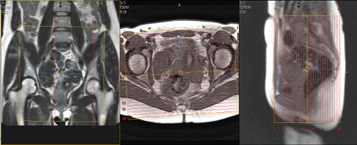
Pelvic floor mri protocol.

Magnetic Resonance Imaging Of The Female Pelvic Floor Radiology Key

Pin On Health
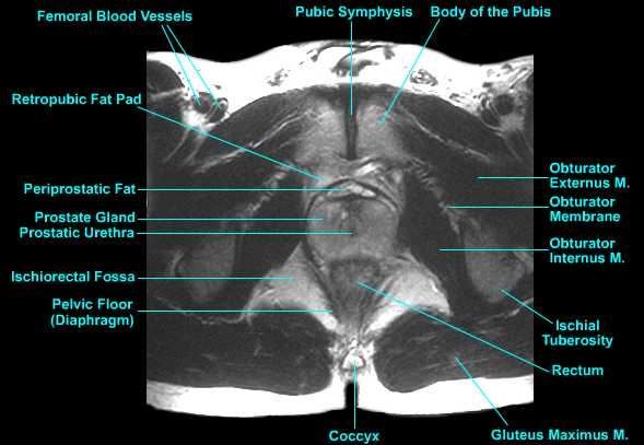
Mri Protocols Mri Anatomy Of The Prostrate

Radiology Rounds Radiology Rounds Medical Specialties Medical Training Radiology

Mr Defecating Proctography Radiology Reference Article Radiopaedia Org

Downloads Kathe Wallace Physical Therapy 1 Physical Therapy Rehabilitation Therapy Therapy

Suggested Protocol For Dynamic Mri Of Pelvic Floor Dysfunction Download Table

Shoulder Imaging Body Bones Image Body
Dynamic Magnetic Resonance Imaging Of The Female Pelvic Floor A Pictorial Review Springerlink

Trigger Point Therapy Quadratus Femoris Trigger Point Therapy Trigger Points Therapy

Pin En Neurologia
Mri Defecography Springerlink

Image Result For Meckel S Cave Cave
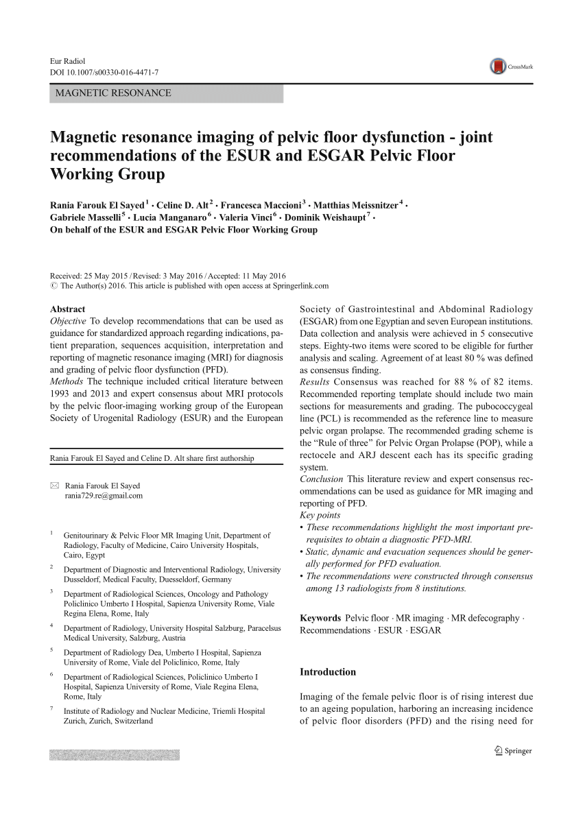
Pdf Magnetic Resonance Imaging Of Pelvic Floor Dysfunction Joint Recommendations Of The Esur And Esgar Pelvic Floor Working Group

Systemic And Visceral Collaterals In 2020 Medical Illustration Interventional Radiology Arteries Anatomy

We Ve Created An Infographic To Explain Embolization Step By Step Check It Out And Let Us Know What You Think Ft Uterine Fibroids Fibroids Fibroids Treatment

Malf Uterinas Medical Ultrasound Diagnostic Medical Sonography Ultrasound Sonography

Non Contrast Pelvic Magnetic Resonance Mr Protocol For Imaging Of Complex Perianal Fistulizing Disease Sages Abstract Archives
Https Encrypted Tbn0 Gstatic Com Images Q Tbn 3aand9gcqtckb4pgywdmdgwt88g2bnhxwz9bowq5l1o0gybunzo Ouy7wl Usqp Cau

Pin On Cervical Injection Exhibits

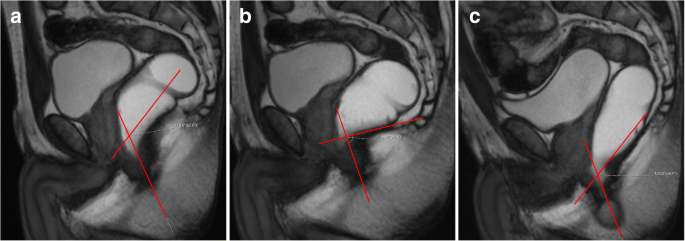
Dynamic Pelvic Floor Mri

Pdf The Urogynecological Side Of Pelvic Floor Mri The Clinician S Needs And The Radiologist S Role

F I T By Jordan Subchorionic Hematoma Cervix Small Circle

Ultrasound Of Callosal Agenesis Ultrasound Corpus Callosum Ultrasound Sonography
Source : pinterest.com
